By Dr. Gregory Mark
All patients have unique cases with various solutions, but some have the opportunity to boost their confidence without worrisome procedures. This treatment applied to one patient in specific. This patient presented a failing bridge between teeth #29-31 due to secondary decay. She was an average dental patient, having medical history within normal limits, and a dental history revealing multiple restorations.
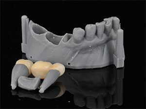
The ideal treatment plan for this patient was to remove the existing bridge, remove the secondary decay, and fabricate a new zirconia bridge. The step-by-step procedure was done using local anesthesia. First, the existing bridge made of PFM (Porcelain Fused Metal) was removed and the secondary decay was excavated. In order to prepare for the crowns, a CEREC Omnicam was used to scan the patient and forward the digital imaging file via CEREC Connect to the InLab software. As we all know, restorations need manual touch and precise materials to accentuate anatomy. Therefore, the crowns were developed using KATANA STML Zirconia for the final restorative material for several reasons: this restorative material had a near identical shade, exemplified the needed strength, and was extremely easy to work with.
 |
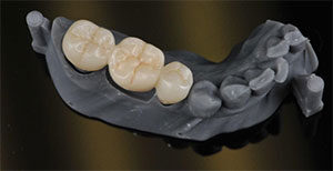 |
| Figure 1 Removable dies and model digitally designed and printed. | Figure 2 Secondary occlusal anatomy carved prior to sintering and staining |
 |
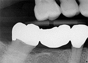 |
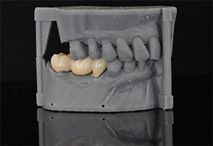
| Figure 3 Stain and glaze was utilized and additionally a diamond paste to polish the bridge | Figure 4 BW post cementation. |
 |
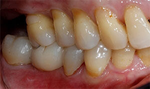 |
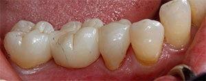
| Figure 5 Buccal view post cementation. | Figure 6 Buccal view post cementation. |
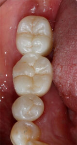 |
| Figure 7 Occlusal view post cementation. |
With this material, the InLab software designed a bridge milled with an MCX5 milling unit. When the final product bridge was milled, it was placed in a Vita Zirconia oven to sinter for 8 hours. In order to bring more vitality to the restoration, and stain and glaze was utilized and additionally a diamond paste to polish the bridge.
The patient returned the following week for the final steps of the procedure. The temporary bridge was removed, and the final cementation of the restoration was conducted. After the fitting, the patient examined the esthetic look and occlusal feel, extremely satisfied with the results. Finally, x-rays and photos were taken, and an overall happier patient was achieved.
Dr. Gregory Mark is a skillful dentist who has been perfecting his art of dentistry since the year 1988. Dr. Mark began his dental career after graduating from Tashkent Medical University in Uzbekistan. Shortly after immigrating to the USA with his wife and two young kids, Dr. Mark excelled at continuing his dental education. He completed an additional three years at NYU College of Dentistry, graduating with honors in 1995. Just a year later, Dr. Mark opened his first private practice. But finally after a decade of devotion to the practice, Dr. Mark decided to expand and relocate to a new, beautifully renovated office in the heart of Forest Hills. The office is equipped with the most advanced technologies; digital imaging, computerized education for the patients, etc. Dr. Mark keeps the office up to date with the latest advancements in the field of dentistry. He is able to do so by studying new techniques learned through various courses taught by the prominent Kois Center, and incorporating them into perfecting his practice. Over the years, Dr. Mark has grown internationally as a mentor for Kois and other major dental teaching corporations. Through his teachings and passion for the field, Dr. Mark has gained recognition for his extensive work beyond basic dentistry.